Symptoms of Age-Related Macular Degeneration (AMD) and How It Is Diagnosed
As you may not notice any signs of age-related macular degeneration, the best way to protect your sight from AMD is to have regular annual eye examinations. During this examination, an ophthalmologist or optometrist will be able to assess your retina is health and if you have any signs of AMD by doing a comprehensive medical eye examination. A condition called Late-Onset Retinal Degeneration (L-ORD) is often mistaken for regular macular degeneration but, in its severest state, affects both central and peripheral vision.
By Lylas G. Mogk, M.D., Edited by Sefy Paulose, M.D., March 2022
A Health and Medication History
- Your overall heath
- Questions about any other risk factors such as smoking, high blood pressure, obesity, etc
Visual Acuity Testing
- Distance and near vision acuity tests to determine the sharpness or clarity of your reading and distance vision
- Testing your vision with different lenses (sometimes contained in a machine called a phoropter) to determine if your vision can be improved or corrected with regular glasses or contact lenses
An Eye Health Evaluation
- Confrontation with visual fields
- Amsler Grid is a black and white grid with a small black dot at its center. Your doctor will have you wear your glasses as you normally would to read, then hold the gride about one foot away from your face. They will ask you to cover one eye and look directly at the center of the dot. While looking directly at the center of the dot and using only your peripheral vision, your doctor will ask you if all the grid lines look straight or if any lines look blurry, wavy or dark. The same will be done for your other eye. Find out how to use an Amsler Grid to detect changes in your vision related to AMD.
- A special microscope, called a slit lamp, is used to examine the anterior segment of the eye (front third of the eyeball), including the cornea, pupil, iris, lens, and aqueous drainage structures.
- A dilated eye (or fundus) examination that can be achieved with the use of special lenses will allow your doctor to see inside your eye and examine the retina. Your doctor might use eye drops to see the retina and optic nerve more clearly. During this exam, the doctor will examine your macula, or the center part of your vision, to see any signs of AMD. You will then be asked to look in different directions so your doctor can examine every section of your retina to assess for other signs of retinopathy.
OCT
- As discussed, your doctor can subjectively look at your macula to assess for any signs of AMD. However, your doctor may want to assess this objectively with a machine that takes a cross-sectional photo of your macula. This test is done through a non-invasive imaging test called Optical Coherence Tomography, or OCT.
- The technician or ocular photographer will have you rest your head on a chinrest and look straight at a target. The machine will then focus on your macula and scan the area to see all the layers of the macula and assess for any changes. Your doctor will be able to view these results and assess the progression of AMD by comparing these results with any past or future test results.
The Amsler Grid
by Audrey Demmitt, R.N.
How Does Age-Related Macular Degeneration (AMD) Damage the Retina?
AMD affects the center portion of the retina, called the macula. This area helps us see up-close and in detail like when we read, thread a needle, or recognize a friend’s face. The macula is damaged by fatty deposits of drusen and thins out in the dry form of AMD. In wet AMD, the damage is caused by fluids and blood leaking under the retina, forming blisters. If left untreated, retinal scarring occurs and vision can be lost permanently. These changes in the macula will cause distortions like blurriness, white, gray, or black spots, and wavy or broken lines in the field of vision. The Amsler Grid can help pick up these distortions and monitor changes in your vision. There are treatments that can dry out the fluid and stop the leaking and preserve vision if received in time.
Explanation of the Amsler Grid
Once age-related macular degeneration has been diagnosed, your eye care professional may give you an Amsler Grid to use at home as an early warning system for changes in age-related macular degeneration, particularly a change from dry to wet AMD. The macula is particularly sensitive to horizontal and vertical lines; therefore, waviness, distortion, or missing lines on the grid may be noticed before a change in visual acuity. This black and white grid looks like graph paper with a small black dot at its center. AMD is a progressive disease that causes gradual visual changes that may not be noticed. This simple testing tool can effectively detect visual changes caused by AMD.
The first image below shows an Amsler Grid as seen with normal vision. The next image is how the Amsler Grid may appear to a person with age-related macular degeneration. These images of the grids are much smaller than normal size so that we can show them to you on this website. If you have been diagnosed with age-related macular degeneration, ask your eye care professional for a real Amsler Grid you can use at home. You can also download a free Amsler Grid from the BrightFocus Foundation or request a magnetic version of the Amsler Grid by calling 1-855-345-6637. Note: this screening test does not replace the need for regular eye exams.
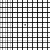 Amsler Grid As Seen with Normal Vision
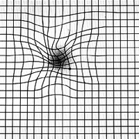 Amsler Grid as Seen with AMD
How To Use An Amsler Grid
The American Academy of Ophthalmology (AAO) recommends people with AMD use the Grid to check their eyesight once a day, every day. Follow these steps:
- Wearing any glasses you normally use to read, hold the grid 12 to 15 inches away from your face in good light.
- Cover one eye.
- Look directly at the center dot with your uncovered eye and keep your eye focused on it.
- While looking directly at the center dot, notice in your side vision if all grid lines look straight or if any lines or areas look blurry, wavy, dark, or blank.
- Follow the same steps with the other eye.
- The first time you look at an Amsler Grid, take note of any irregularities in the grid and where they appear in the four quadrants. Think of this as your baseline. Now if any changes occur, contact your retinal specialist immediately.
This simple daily routine will take less than a minute and can be sight-saving. Remember: Early detection of vision changes means timely treatment that can save your vision. To find out more, Watch this video by the AAO on how to use an Amsler Grid.
Reference
Have AMD? Save Your Sight with an Amsler Grid – American Academy of Ophthalmology (aao.org)