Refractive Error and Astigmatism
The power of your eye to focus and see an image is dependent on several structures within the eye:
- The cornea is a transparent dome-shaped tissue that forms the front part of the eye. It functions as a window and allows light to enter the eye. It also begins focusing light rays that allow you to see words and images clearly.
- The lens comprises transparent, flexible tissue directly behind the iris and the pupil. It is the second part of the eye, after the cornea, which helps to focus light and images on the retina. Because the lens is flexible and elastic, it can change its curved shape to focus on objects and people nearby or at a distance.
- To see as clearly as possible, images must be focused by the cornea and lens directly onto the retina, the light-sensitive tissue that lines the inside surface of the eye, much like the film in a camera. The optic nerve transmits visual information from the retina to the brain.
- For vision to be as sharp as possible, the focusing power of the lens and cornea must correspond appropriately to the length of the eye so that the light rays ultimately come together at the retina. If they focus either in front of the retina or behind it, the image will not be as clear as possible, and there will be a refractive error.
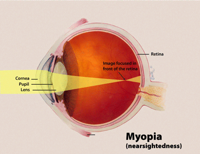
Myopia or Near-Sightedness
People with myopia, or near-sightedness, can see close-up objects clearly, but distant objects are blurred.
- In myopia, light rays are brought to focus in front of the retina, i.e., falling short of the back of the eye.
- This occurs either because the focusing power of the cornea and lens is very high because the eyeball is too long from front to back, or both.
 Source: National Eye Institute
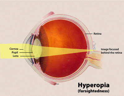
Hyperopia or Far-Sightedness
In contrast, people with hyperopia, or far-sightedness, can see distant objects clearly, but close-up objects are blurred.
- In hyperopia, light rays are brought to focus behind the retina.
- This occurs either because the focusing power of the cornea and lens is very low. After all, the eyeball is short in length from front to back or both.
 Source: National Eye Institute
How Is Refractive Error Diagnosed?
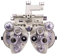
Refractive error is diagnosed by an ophthalmologist or optometrist by using a machine called a phoropter (pictured at left), which is placed in front of the patient’s eyes to trial, or evaluate, various strengths of eyeglass prescriptions.
The patient participates by informing the doctor which of the various eyeglass strengths provides the clearest image, which allows the doctor to fine-tune the prescription until the final correct prescription is reached.
In patients who cannot provide the necessary feedback (including people with physical and cognitive disabilities and very young children), the doctor can assess the refractive error via a process called retinoscopy. To do a retinoscopy, the doctor uses a device called a retinoscope to shine a light into the patient’s eye. The doctor then trials the various lenses while observing the light reflection, or reflex, in the patient’s eye to determine the correct prescription strength.
How Is Refractive Error Treated?
Refractive error is generally corrected by glasses or contact lenses that help focus the image correctly on the retina. There are also a variety of refractive laser surgeries. Most of these work by changing the curvature, and thus power, of the cornea. These include the following:
- photorefractive keratectomy (PRK)
- laser in-situ keratomileusis (LASIK)
- laser epithelial keratomileusis (LASEK)
- epiLASIK
Corneal incision procedures, such as radial keratotomy or limbal relaxing incisions, involve placing incisions into the cornea to treat myopia or astigmatism.
Additional refractive surgeries include the placement of intrastromal corneal ring segments (called Intacs) into the cornea to treat low degrees of myopia or a phakic intraocular lens, in which an implantable lens is inserted in the eye to treat moderate to high myopia. These options should carefully be discussed with a refractive surgeon to weigh the risks and benefits and to choose to most appropriate procedure if refractive surgery is pursued.
If I Do Not Wear My Glasses, Will My Eyes Get Worse?
Unless the person is young enough that the visual pathways are still developing (see Amblyopia), neither wearing nor not wearing eyeglasses will alter the health or visual potential of the eye.
- For an adult, failure to wear eyeglasses for distance will just mean that you won’t see as clearly when you’re not wearing them, but is will not worsen your vision or lead to requiring stronger glasses.
What Is Astigmatism?
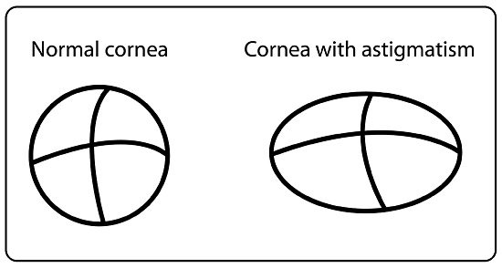
Astigmatism is a blurring of vision due to an irregular shape in the cornea or lens. In eyes without astigmatism, the cornea and lens have a more or less similar curvature in all directions. This allows light to be focused to a single point on the retina.
People with astigmatism have more curvature in one direction, or meridian, than in another (see Figure below), so that light is not able to focus on a single point on the retina. This results in blurring of vision at all distances. Normally, the cornea’s surface is rounded, much like a basketball; with astigmatism, however, the cornea is shaped more like an American football.
 Source: National Eye Institute
In the vast majority of cases, astigmatism is a result of the shape of the cornea (and sometimes the lens) that is present since birth.
However, there are certain eye conditions which are associated with astigmatism, such as keratoconus, corneal lesions, scars, prior surgery in the cornea, and a number of other diseases.
Astigmatism is usually accompanied by myopia or hyperopia. The degree of astigmatism can be determined during the refraction portion of an eye examination. A refraction helps measure the sharpness or clarity of both close-up and distance vision by testing with different lenses to determine if vision can be improved or corrected with regular eyeglasses or contact lenses.
In addition, an automated or manual machine called a keratometer can determine the cornea’s curvature. Corneal topography can map out the entire cornea and its curvature.
How Is Astigmatism Treated?
People with mild astigmatism may not notice significant blurring and may elect to forgo correction of their astigmatism. In contrast, people whose vision is affected by astigmatism can benefit from an astigmatic correction in their glasses or contact lenses.
Astigmatism can also be corrected by the refractive surgeries discussed previously, in which incisions or lasers alter the corneal curvature to alleviate astigmatism.
In addition, patients with moderate astigmatism undergoing cataract surgery may benefit from selecting an intraocular lens implant, called a toric lens, that corrects for astigmatism.
By Mrinali Patel Gupta, M.D. Updated by Lylas Mogk, M.D. 9/23