Cataract Surgery and Risks
Article by Tina D. Turner, M.D. and updated by Sefy Paulose, M.D., March 2022
Treatment: Surgical Removal
The permanent fix for cataracts is to remove the cataract surgically. To date, no medications or eye drops have been proven to reverse cataract formation. However, your doctor may see if changing your glasses prescription will help you obtain better vision. This is because cataracts can cause nearsightedness. However, the only treatment for a cataract is the surgical removal of the natural lens.
When to Remove?
Most age-related cataracts are a normal part of aging, so simply “having” a cataract should not mean it should be removed. Many people with cataracts do not have any visual symptoms. If you are told you have a cataract, but it does not interfere with your activities of daily life or prevent you from leading an active and productive life, then your doctor may tell you the cataract can be monitored. However, if you are experiencing difficulty reading, disabling glare while driving or difficulty engaging in your normal life activities, it may be time to consider cataract surgery. In summary, the right time to remove a cataract depends on the patient’s symptoms. However, if an individual has cataracts in both eyes that require surgeries, these surgeries will usually be performed a few weeks apart. Cataract surgery on both eyes simultaneously is not recommended because there is a possibility of complications affecting both eyes; the most worrisome is infection.
Professor John Hull (1935-2015), Author of Touching the Rock: An Experience of Blindness
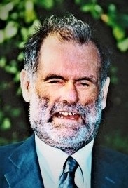
The late Professor John Hull was the author of Touching the Rock: An Experience of Blindness, his compelling memoir that documents the process of becoming blind. As a young university lecturer in the early ’60s, Hull had adapted to cataracts and the early signs of retinal detachment brought on by numerous surgeries. “For the first few years after I registered as being blind,” he said, “I was not, in effect, a blind person. I was a sighted person who couldn’t see. It’s such a difference. It wasn’t until the light sensation completely vanished and I knew there was no way back that I said, ‘I’ve got to try to understand blindness; otherwise it will destroy my life.’” Learn more about ways to find emotional support for you – and your family members – after a vision loss diagnosis:
How Much Should the Cataract Develop Before Having Surgery?
A cataract does not have to become “ripe” before it can be removed. In the past, the lens could not be extracted safely from the eye unless it was at a relatively advanced stage of development. With modern advances in cataract surgery, the lens can now be removed from the eye at any stage of development.
The Patient’s Decision
It’s important to understand that it is the patient who should – and must – decide to undergo cataract surgery. The doctor’s responsible for educating patients and giving them the knowledge they need to make an independent and well-informed decision regarding cataract treatment.
Anesthesia and Medication
Commonly, cataract surgery is performed with topical anesthesia. This is accomplished by instilling a powerful numbing medication into the eye. It is usually accompanied intravenously by medication in the patient’s arm to help them feel relaxed and comfortable. This is the least risky form of anesthesia, and most patients do extremely well with topical anesthesia and some intravenous sedation. Sometimes, medication is injected around the eye socket to numb the eye and paralyze eye and eyelid movement. However, these injections carry their own risk and are being used less frequently.
General Anesthesia
Sometimes, general anesthesia may be needed. Since cataract surgery performed with topical anesthesia requires patient awareness and cooperation, general anesthesia is usually required for children, patients with developmental delays, and patients with dementia. During cataract surgery, patients must lay flat and still; therefore, patients with movement disorders, such as Parkinson’s Disease or restless leg syndrome, may also require general anesthesia. Patients who have difficulty breathing while lying flat, or who have back or neck pain/disorders that prevent them from being comfortable when lying flat may also require general anesthesia for cataract surgery.
Discuss Your Options
After deciding to have cataract surgery, the patient and physician should discuss the options for correcting vision post-surgery. Artificial lenses, implanted in the eye during cataract surgery to replace the natural lens being removed, can make the vision clear once again and, in some cases (but not always), reduce the need for corrective eyeglasses after surgery. The surgeon will take special eye measurements before surgery, including the eye’s length and the cornea’s curvature, to determine what power the artificial lens should be. Cataract surgery can decrease an individual’s dependency on eyeglasses and sometimes eliminate the need for eyeglasses after surgery. However, some patients will still need eyeglasses to correct their distance and/or reading vision to 20/20.
Performing Cataract Surgery
Most cataracts are highly treatable. Cataract surgery is one of the most common surgeries performed in the United States, with approximately 98% of patients experiencing improved vision if no other eye conditions are present.
Two very small incisions (one larger, approximately three millimeters, or one-tenth of an inch, and one smaller, approximately one millimeter, or one thirty-second of an inch) are made in the cornea, which is the transparent dome-shaped tissue that covers the front part of the eye. A viscous (thick, sticky, glue-like) material is injected into the front part of the eye to help maintain its shape during surgery. This viscous material is made from substances that occur naturally in the body. Because it is thick, this material will not leak out of the incisions during surgery.
According to the National Eye Institute:
You can get cataracts in one eye or both eyes — but they can’t spread from one eye to the other.
By age 80, most people either have cataracts or have had cataract surgery.
Cataract surgery is one of the most common operations in the United States.
Phacoemulsification
Phacoemulsification was introduced more than 40 years ago and is now the most common surgical method used to remove cataracts. The surgeon creates an opening in the natural “sac” or “bag” that holds the lens in place, called the lens capsule. The lens is separated from the lens capsule by using a balanced salt solution.
Once the capsule is open and the lens can move freely inside it, a special ultrasound device is used to break the lens into small pieces and suck it out of the eye. This technique is called phacoemulsification.
Prior to the development of phacoemulsification, the lens used to be removed in one solid piece through a very large incision (8-12 millimeters). That surgery entailed considerably more risk and had a significantly longer recovery time.
After removing the lens, additional viscous material is injected into the capsule to hold it open and make room for the new artificial lens. The folded artificial lens is inserted into the “sac” or capsule, allowing it to unfold. The viscous material that maintained the shape of the eye during surgery is removed. The two incisions usually self-seal and do not require stitches.
Femtosecond Laser for Cataract Surgery, or Laser-Assisted Cataract Surgery
Femtosecond lasers have been used in ophthalmic surgery since 2001, and in the late 2000s, work began on their use in cataract surgery. In 2008, the first laser-assisted cataract surgery was performed in Hungary. After gaining FDA approval, the first laser-assisted cataract surgery was performed in the United States in 2010. Since that time, it has been gaining acceptance and popularity.
The laser does not take the place of manual cataract surgery. As stated, the laser “assists” in removing the cataract; phacoemulsification is still used to remove the cataract itself.
The laser performs three key steps in the cataract surgery procedure:
- the corneal incisions
- opening of the capsule containing the cataract
- the initial sectioning of the cataract into smaller pieces.
It performs these three steps with incredible precision, and this aspect may prove it to be superior to the current technique in which the surgeon manually performs these steps. The laser can also be used to make incisions in the cornea to treat certain types and amounts of astigmatism.
Further data and well-designed studies are needed to prove that this method is associated with better outcomes and fewer complications than phacoemulsification alone, and much work is being done on this front.
Because insurance does not cover the cost of laser-assisted cataract surgery, patients must contribute a significant out-of-pocket payment. It is important to discuss with your physician what type of procedure would be best for you.
Find out more about this type of surgery.
How Long Is the Recovery Time After Cataract Surgery?
Some patients see very well the day after cataract surgery. Other patients see well a few days after surgery, and still others may need a full month to reach their maximum vision improvement.
The First Week after Surgery
During the first week after surgery, it generally is recommended that the patient keep his or her eye covered at all times, either with eyeglasses or an eye shield, to protect it from being bumped or rubbed. A small amount of pressure can easily open the incision, and protecting the eye prevents this.
Also, it is recommended that the patient refrains from (a) bending with the head below the waist, (b) lifting more than 10 pounds, and (c) straining (on the toilet, for example) to the point of holding one’s breath. These activities increase the pressure inside the eye and can open the incision.
Antibiotic and anti-inflammatory eye drops are used weeks after cataract surgery to help prevent infection and control inflammation.
A Cataract Cannot Come Back After Surgery
This is because cataract surgery permanently removes the lens and places an artificial lens in the eye. Unlike our natural lenses, these artificial lenses do not harden, yellow or cloud over time. However, the lens capsule, the small “sac” or membrane that once surrounded the natural lens and held it in place, can change.
Capsular Opacification
The lens capsule is like the shell of a peanut M&M. Over time, it can grow a small membrane which can seem like the cloudiness of the artificial lens itself. This is called capsular opacification, which develops in approximately 25% of patients after cataract surgery. If this occurs, the patient may develop symptoms similar to those of a cataract, such as blurry or hazy vision, difficulty reading regular print, and sensitivity to bright lights and glare. Posterior capsular opacification is treated with a laser to create an opening in the center of that membrane to allow light to enter the eye. The procedure is painless, requires less than five minutes, and is usually performed in the doctor’s office.